
Christian Mata defends his PhD thesis “Web-based Application for Medical Imaging Management”
The rapid growth of digital technology in medical fields during the last few years has increased the need for applications able to manage patient medical records, imaging tests, and chart information. Technologies related with digital database architectures, storage and transmission protocols, management of large volumes of data, and security are being improved every day, allowing the possibility of reading, analyzing, and even diagnosing a case far from the medical center where the information was acquired.
Furthermore, the inclusion of web-based applications applied to the medical imaging field has growing demand over the last few years in the medical community. The study of how applications can store and exchange this huge amount of information is becoming a new paradigm to solve present day limitations to managing medical data. Indexing tools based on the standard protocols allows a fast retrieval of the information and securities issues. Web-based applications in computational medicine have become increasingly important over the last few years. It supposed a new paradigm in the telemedicine and e-Health areas in order to assist and enhance the prevention, diagnosis and treatment of ailments. Furthermore, the training of radiologists and the management of medical databases are also becoming increasingly important issues in the field.
In this domain, firstly, this thesis analyzes the main advantages and drawbacks of the reviewed work in web-based applications applied to the medical imaging field. Then, a web-based medical application including a system architecture based on digital imaging is presented. One of the main purposes of this PhD thesis is to offer an easy accessibility to web-based applications for users using a new system architecture. They can connect from any part of the world, providing this to be a time-saving and efficient mechanism to find information. In fact, this information can be saved directly in computers without the tiring task of searching in libraries or alternative databases. However, the access of the previous process is also available with the possibility of downloading medical annotations using query forms. Then collaborative work is possible by considering several opinions from different experts. Our system, being dedicated to medical imaging, manages the corresponding specific
Digital Imaging and Communications in Medicine (DICOM) format. Finally, our web-based application also provides a high level security system.
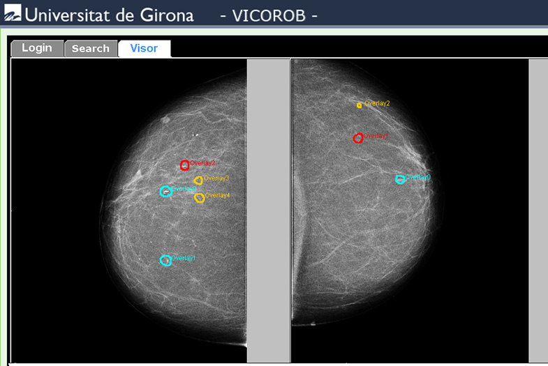
Prostate and breast cancer are the most common cause of cancers in men and women, respectively. Medical imaging plays an important role in breast and prostate cancer detection and evaluation. Then to prove that our web-based medical application could be applied in different medical disciplines, the main part of this thesis is the implementation of two frameworks as a Java applet interface designed as a web-based tool in the domains of mammography from X-rays in radiology, and of prostate imaging from an Magnetic Resonance Imaging (MRI). This aims to facilitate the diagnosis of new mammographic and prostate cases by providing a set of image processing tools that allow a better visualization of the images, and a set of drawing tools used to annotate the suspicious regions (overlays). Each annotation allows including the attributes considered by the experts when issuing the final diagnosis. The overall set of overlays is stored in a database as eXtensible Markup Language (XML) files associated with the original images. Finally, an exhaustive evaluation of the results is also discussed in this thesis. For the application on mammography, the experimental study is performed in order to evaluate the scalability, complexity and response speed at the proposed tool. For the application on MRI of prostate cancer, the evaluation focused on the decrease of the variability of the expert assessments when collaborative work is performed. To conclude, a new architecture with the main goal of managing patient databases with potentially multi-modal imaging is presented such as for an MRI of the prostate cancer and evaluation from potentially several experts.
_____________________________________________________________
El ràpid creixement de la tecnologia digital en el camp mèdic ha generat la necessitat de desenvolupar aplicacions que siguin capaces de gestionar grans volums de dades. Conseqüentment, les bases de dades digitals, els protocols d’emmagatzement, la transmissió i seguretat es van millorant cada dia possibilitant la lectura, anàlisi i gestió de diagnòstics clínics tant en el mateix centre mèdic o externament.
La inclusió d’aplicacions web aplicades a la pràctica mèdica s’ha incrementat notablement mitjançant els avenços tecnològics dedicats a l’anàlisi i processat d’imatges digitals. En aquest sentit, el camp de la imatge mèdica te un rol molt important en aquesta última dècada gràcies a eines basades en protocols que compleixen un format estàndard i que permeten una ràpida recuperació de la informació. Malgrat això, un dels majors inconvenients és l’intercanvi i emmagatzematge de gran quantitat d’informació provinent de centres mèdics. Ha suposat sens dubte un nou paradigma en el camp de la telemedicina en trobar aplicacions que puguin ajudar i millorar la prevenció, diagnòstic i tractament de pacients. Així com la formació de radiòlegs i l’aprenentatge per la gestió de base de dades mèdiques cada vegada és més necessària en el camp mèdic.
Hi ha molts avantatges en l’ús d’aplicacions web en l’àmbit mèdic. La principal avantatge es deu a la fàcil accessibilitat per als usuaris que poden connectar-se des de qualsevol lloc del món, l’estalvi de temps i l’eficient mecanisme per la gestió de la informació. A més, proporcionen eines que faciliten la descàrrega d’anotacions mediques així com alts nivells de sistema de seguretat. En el camp mèdic és imprescindible tenir sistemes de seguretat i protecció de la informació dels pacients. Per tant, les tecnologies d’imatges digitals s’han convertit beneficioses per les pràctiques mèdiques modernes i sistemes d’atenció de salut proporcionant ajuda per al diagnòstic, el tractament i la cirurgia.
Durant els últims anys, la taxa d’incidència en la detecció de càncer s’ha reduit lleugerament en els homes i s’ha mantingut estable en les dones, mentre que les taxes de mortalitat per càncer es va reduir en un 1,8% anual en els homes i un 1,4% per any en les dones. Aquesta reducció es deu en gran part a l’anàlisi i processament d’imatges mèdiques i tot aquest conjunt d’aplicacions d’ajuda al diagnòstic del càncer. D’una forma molt notòria contribueix a la seva detecció precoç mitjançant cribratge, diagnòstic, biòpsia guiada per imatges i arquitectures per la gestió de dades mèdiques tals com base de dades d’imatges digitals. En realitat, els càncers de pròstata i mama són la causa més comú de mort per càncer comparat amb els altres. Per aquest motiu, el principal objectiu és millorar les eines i aplicacions actuals per proporcionar una millor instrumentació per obtenir una millor qualitat d’imatges, així com, metodologies i procediments amb la finalitat de millorar la interpretació de les lectures de la imatge.
En aquesta tesi es realitza una revisió bibliogràfica de les principals publicacions recents en els últims anys en aplicacions mèdiques basades en web. Aquest estudi analitza els avantatges i inconvenients dels treballs d’investigació en el camp de la imatge mèdica, així com les arquitectures de base de dades per a la gestió d’imatges digitals. La part principal d’aquesta tesi és la implementació d’una eina basada en la web amb la finalitat de demostrar la integritat i aplicació en diferents disciplines mediques. En aquest sentit, l’aplicació proposada en aquest projecte de tesis ha sigut implementada com a eina d’ajuda al diagnòstic de càncer de mama i pròstata. L’objectiu és facilitar el diagnòstic proporcionant un conjunt d’eines de processat d’imatge que permetin una millor visualització de les imatges, i un conjunt d’eines d’anotació de regions sospitoses o malignes (superposicions). Cada anotació permet incloure tots els atributs i especificacions considerades pels experts a l’emetre el diagnòstic final. S’han dissenyat diferents arquitectures per a la gestió de base de dades (per exemple PACS per emmagatzemar imatges monogràfiques). Per altra banda, el conjunt global d’anotacions s’emmagatzemen en una base de dades d’arxius XML associats a les imatges originals. Conseqüentment, aquesta nova arquitectura es presenta amb l’objectiu d’obtenir una base de dades de casos diagnosticats i validats per radiòlegs experts per a la formació de radiòlegs novells. Finalment, conclusions i noves línies d’investigació associades al projecte com a treball futur són presentades en aquesta tesi.
No Comment