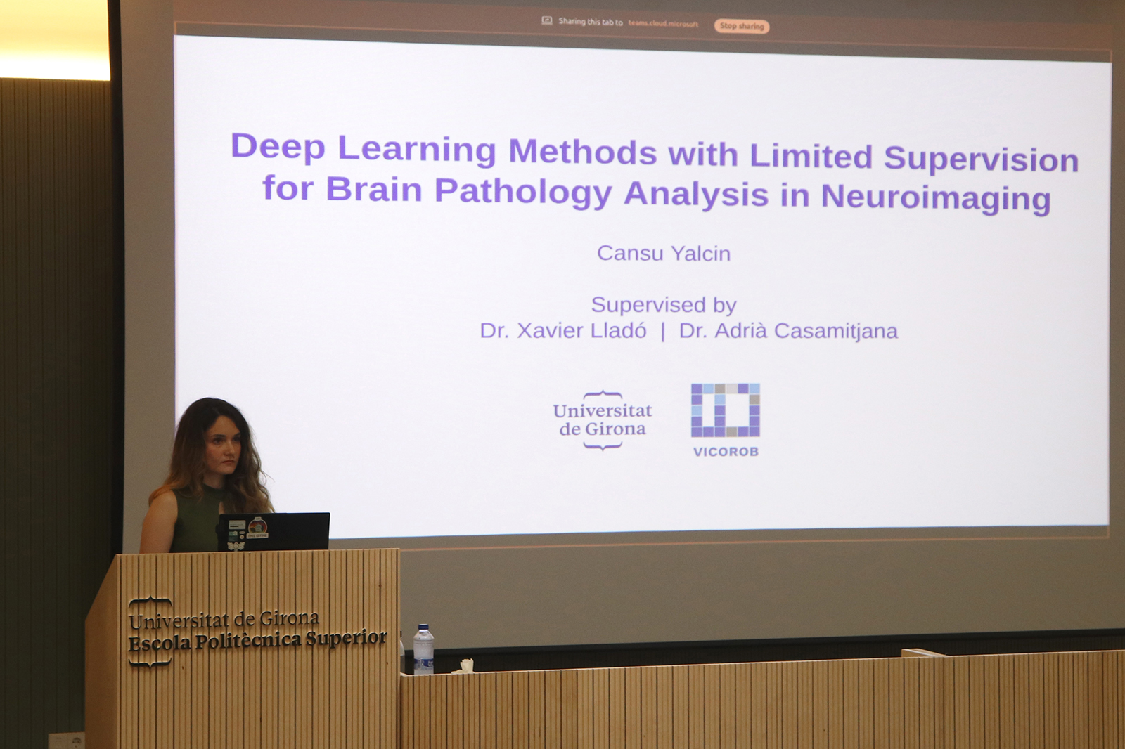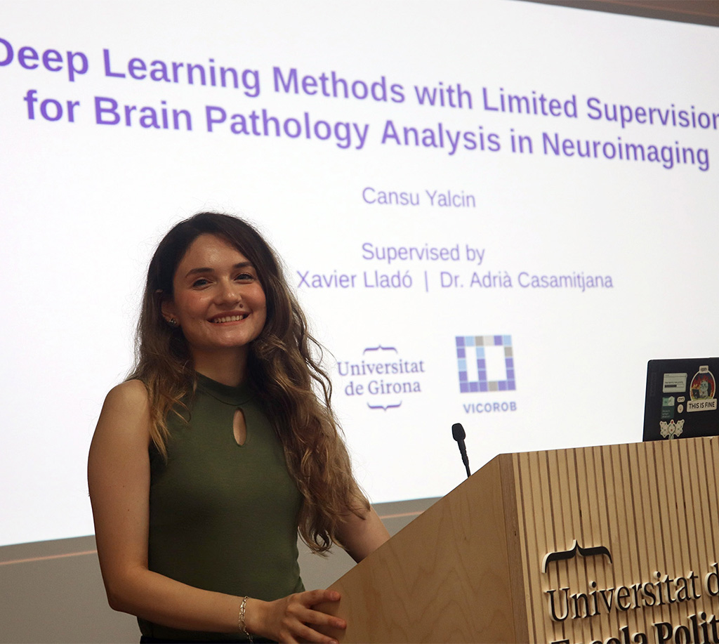
By: Cansu Yalcin
Supervised by: Dr. Xavier Lladó and Dr. Adrià Casamitjana
Thesis Overview
Brain pathology analysis through medical imaging is critical for clinical diagnosis, treatment planning, and patient outcome prediction. Computed tomography (CT) and magnetic resonance imaging (MRI) have become essential imaging modalities for assessing two major classes of brain pathologies: intracranial hemorrhage (ICH) and brain tumors. However, manual interpretation and analysis of these imaging studies are time-consuming, labor-intensive, and susceptible to intra- and interobserver variability. In recent years, artificial intelligence-based methods have shown great potential in automating the detection, segmentation, and quantification of brain lesions, thereby supporting clinical workflows and improving diagnostic accuracy. Nevertheless, these methods typically rely on large amounts of annotated data to achieve high performance. In medical imaging, obtaining such annotations is particularly challenging due to the need for expert knowledge, as well as the significant time and economic costs associated with manual labeling. As a result, while vast amounts of medical imaging data are routinely acquired in clinical practice, only a small fraction is accompanied by high-quality annotations. This imbalance highlights the need for approaches that can effectively leverage limited labeled data, motivating the development of learning strategies under limited supervision.
The main goal of this PhD thesis is to develop robust deep learning methodologies for neuroimaging analysis under limited supervision. To address annotation scarcity, we explore several complementary strategies. In the first stage, we propose a framework for hematoma expansion (HE) prediction in spontaneous intracerebral hemorrhage (ICH) using baseline CT scans. This approach combines a classification model with synthetic CT image generation to enhance robustness and alleviate data limitations. Our results show that integrating synthetic data with conventional augmentation improves both predictive performance and generalization. In the second stage, we develop semi-supervised deep learning frameworks that leverage both labeled and unlabeled data for binary brain lesion segmentation in brain tumors and spontaneous ICHs. We introduce a novel method that combines feature perturbation with mutual learning, achieving significant improvements in low-annotation settings (10–20% labeled data). In the third stage, we extend semi-supervised learning to multi-class hemorrhage segmentation, a more complex task requiring the differentiation of multiple hemorrhage subtypes. We conduct a comprehensive benchmark of established semi-supervised methods within a unified nnUNet framework, providing a systematic evaluation of different strategies for this setting.
All proposed methodologies are evaluated on both institutional datasets from collaborating hospitals and public international benchmarks, enabling objective comparison with state-of-the-art approaches. Notably, this work was carried out in close collaboration with the medical team at Hospital Dr. Josep Trueta, with clinical expertise integrated throughout the research process from problem formulation and data acquisition to result interpretation and validation. By combining deep learning techniques with clinical insight, this thesis aims to deliver practical tools that support clinical decision-making and ultimately improve patient care in brain tumors and ICHs. Emphasis is placed on reproducibility and clinical applicability, with all methods and experimental pipelines made publicly available to facilitate future research and the development of reliable automated decision-support systems.