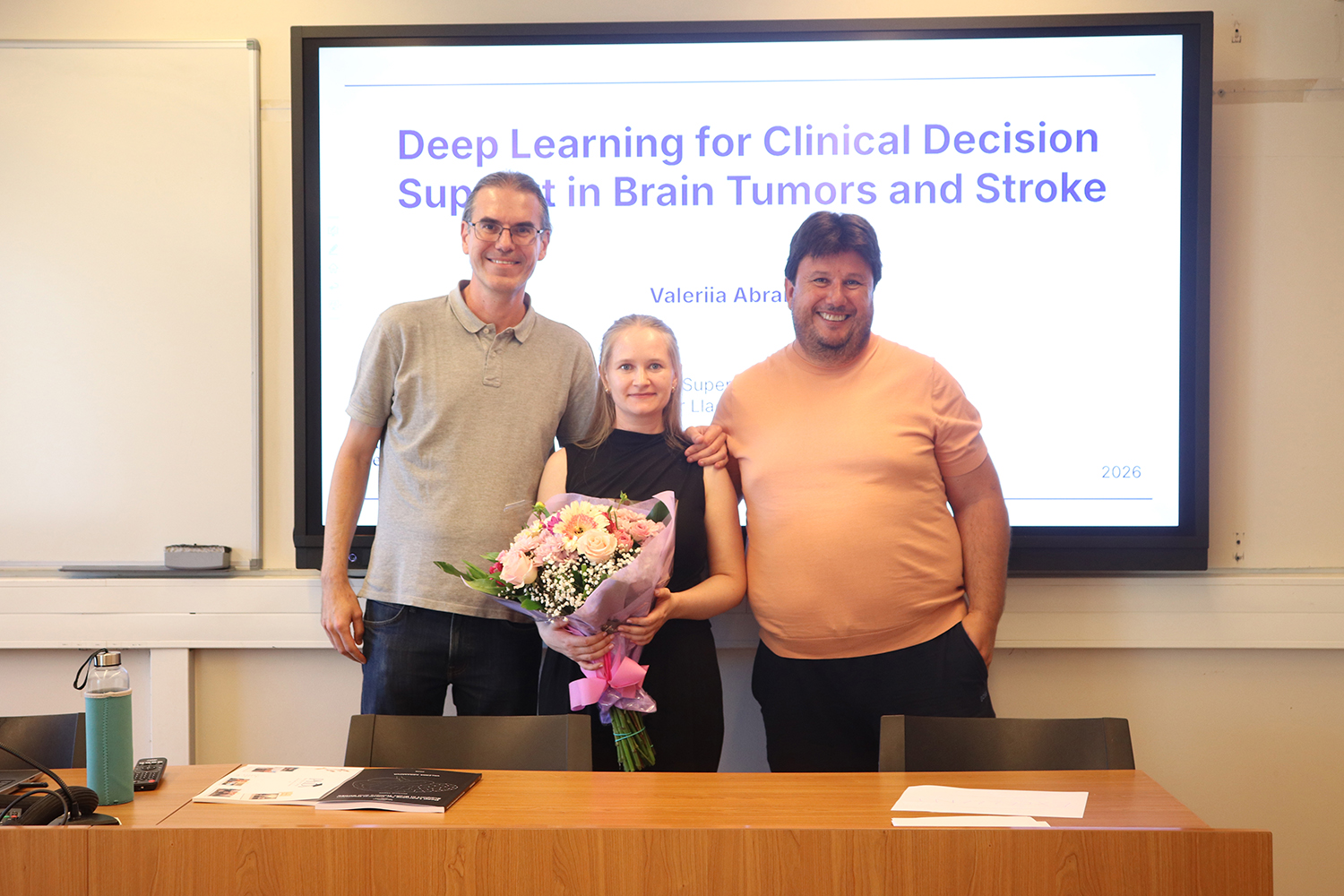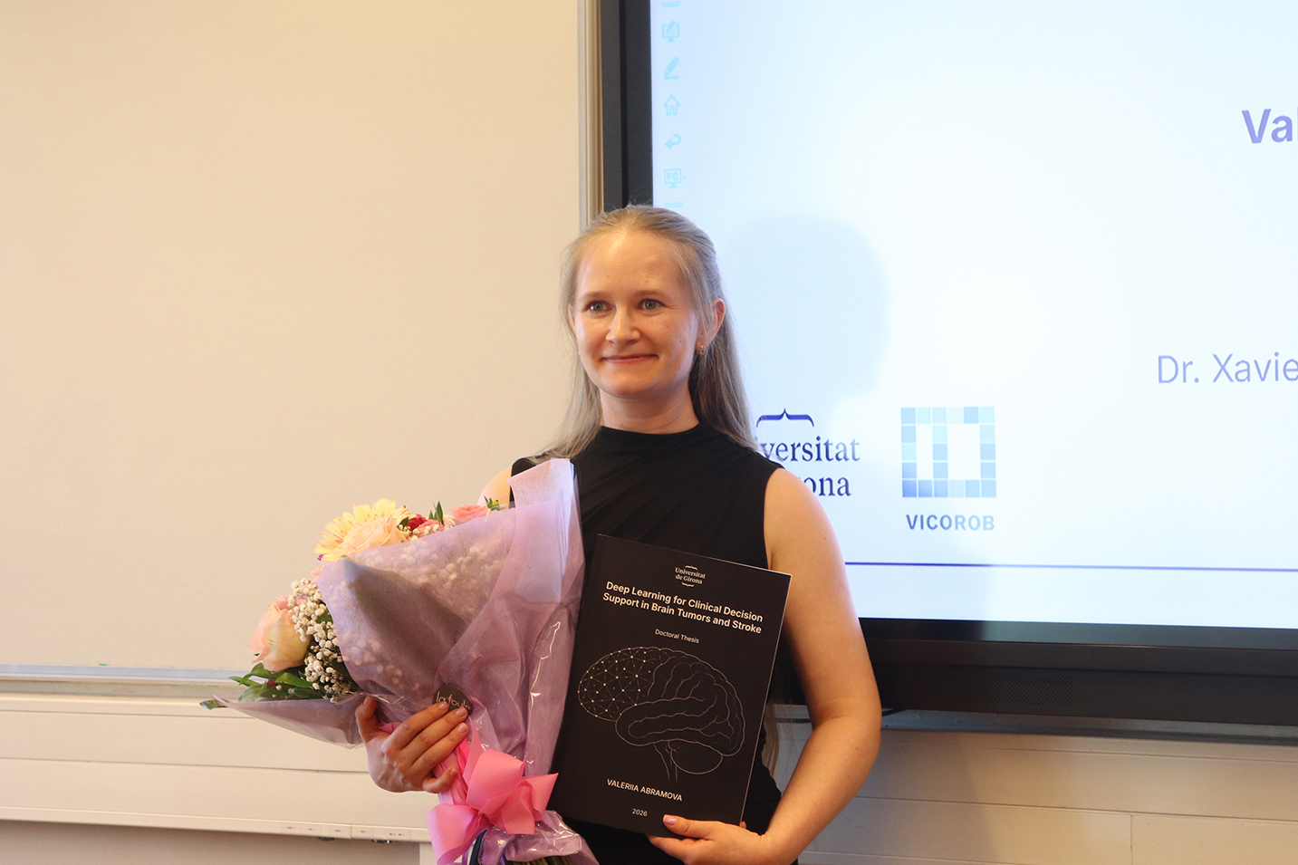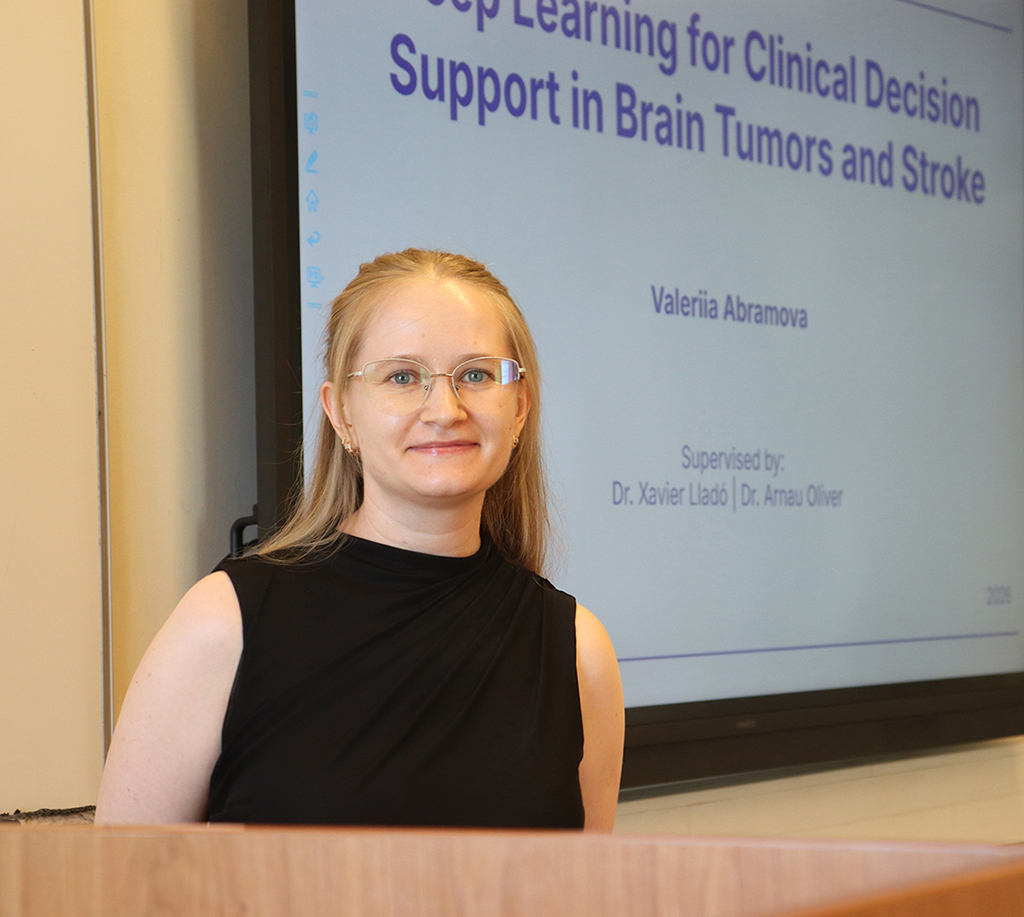
By: Valeriia Abramova
Supervised by: Dr. Xavier Lladó and Dr. Arnau Oliver
Thesis Overview
This PhD thesis focuses on the development of artificial intelligence methods based on deep learning for the analysis of brain images and, ultimately, to help improve the diagnosis and treatment of common neurological diseases such as stroke and meningioma.
Stroke is one of the leading causes of death and disability worldwide. It can be either ischemic or hemorrhagic, and in both cases the diagnosis relies on brain imaging examinations, most commonly computed tomography (CT). Meningioma, on the other hand, is the most common primary brain tumor. It is often detected and monitored through periodic magnetic resonance imaging (MRI) examinations, and when treatment is required, especially radiotherapy, treatment planning is based on these images to accurately define the area to be irradiated. Therefore, medical imaging is a key tool throughout all stages of the clinical management of these diseases.
In this context, this thesis explores how deep learning can make different steps of the clinical workflow more efficient and accurate. First, a method is proposed to predict the possible evolution of intracerebral hemorrhage in patients with hemorrhagic stroke using only the initial available CT scan. One of the main challenges is the limited availability of clinical data, and therefore a strategy is also proposed to generate synthetic examples simulating possible lesion evolutions. These synthetic images are used to train the model and improve its predictive capability, achieving competitive results compared with state-of-the-art methods.
Second, a system is developed to detect large vessel occlusions, one of the main causes of ischemic stroke, from CT angiography images, and to identify the affected vascular segment. The method focuses on a specific anatomical region, the Circle of Willis, which contains the most relevant vessels. This allows the analysis to be simplified, the process to be accelerated, and a high level of reliability to be maintained. The results have been validated using real hospital data and independent test sets, demonstrating good generalization capability.
Third, a method is presented for the segmentation of meningioma tumors in MRI images used for radiotherapy planning. The main contribution is a richer representation of the tumor, which considers not only the lesion itself but also its transition zone. This additional information improves segmentation accuracy and achieves better results, even in comparison with other recent approaches. The method was evaluated and validated in an international challenge, where it achieved first place.
Overall, this thesis demonstrates how artificial intelligence can contribute to making brain image analysis more automated, accurate, and efficient. An important part of the work was carried out in collaboration with the medical team at Hospital Dr. Josep Trueta, integrating real clinical knowledge throughout all stages of the project. The ultimate goal is to provide tools that can have a direct impact on clinical practice and contribute to improving the care of patients with stroke and brain tumors.