By Kaisar Kushibar
Supervised by Dr. Sergi Valverde / Dr. Arnau Oliver / Dr. Xavier Lladó
Abstract
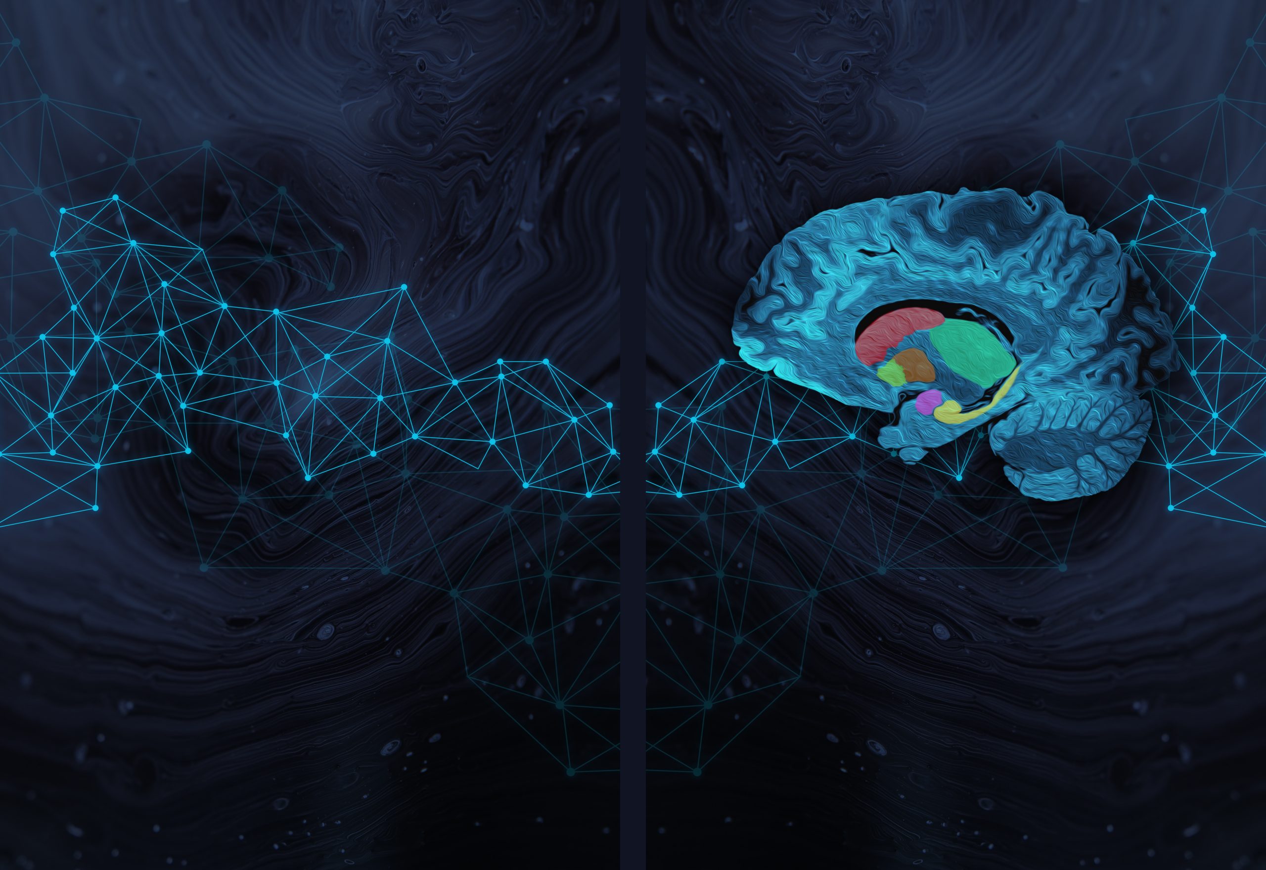
The sub-cortical brain structures are located beneath the cerebral cortex and in- clude the thalamus, caudate, putamen, pallidum, hippocampus, amygdala, and ac- cumbens structures. These bilateral structures – symmetrically located within the left and right hemispheres – are involved in systematic activities such as emotion, pleasure, memory and hormone production. Their deviations in volume are associ- ated with dierent neurological diseases such as Alzheimer’s disorder, bipolar dis- order or multiple sclerosis, among others. Manual segmentation of these structures is a time-consuming task and suers from rater inter- and intra-variability. There- fore, developing automated methods for accurately segmenting the sub-cortical brain structures is important and it is an active research area.
This PhD thesis focuses on the development of deep learning based methods for accurate segmentation of the sub-cortical brain structures from Magnetic Reso- nance Images (MRI). This goal has been carried out in several stages. In the first stage, we have proposed a 2.5D – i.e. three 2D orthogonal planes of a 3D volume – Convolutional Neural Network (CNN) architecture that combines convolutional and spatial features. Additionally, we proposed a selective sample selection technique from structure boundaries. The experimental results demonstrated the eectiveness of the proposed approach in accurately segmenting all the sub-cortical brain struc- tures and has shown state-of-the-art performance on well-known publicly available datasets – Multi-Atlas Labelling Grand Challenge MICCAI 2012 and Internet Brain Segmentation Repository (IBSR).
In general, CNN’s performance drops when tested with images from a dierent domain than the training set. This problem is referred to as the domain shift eect, where a network trained with MRI volumes with the same properties does not generalise to other images with dierent properties such as scanner type, imaging protocol and resolution. In the next stage of this PhD, we addressed this problem of domain adaptation using a supervised transfer learning strategy. We showed the eect of domain shift on the performance of deep learning methods and how our proposal not only resolves this issue but also drastically reduces the number of training images and trainable parameters of the network.
In the following stage, we developed a new approach that did not even require any manually annotated images for domain adaptation – an unsupervised deep learning method. In this approach, we proposed to employ a histogram loss to minimise the eect caused by domain dierences directly within the convolutional layers of the CNN. This approach showed significant (p < 0.001) improvements from baseline segmentation results, where no domain adaptation was applied, and showed compa- rable results to unsupervised FIRST method. In order to show its robustness, we extended our unsupervised domain adaptation method for segmenting brain white matter hyperintensities. The experimental results showed that adapting the network in an unsupervised manner improved the baseline and outperformed traditional un- supervised methods used for this task.
All these completed stages paved the way for achieving an accurate and robust automated deep learning based method for segmenting all the sub-cortical brain structures. Moreover, this PhD thesis has been part of research frameworks within the projects of the ViCOROB group and dierent collaborating hospital centres. Furthermore, the methods developed during this PhD thesis were compiled into a toolbox and made publicly available for the research community.