By: Uma-Maria Lal-Theran Estrada
Supervised by: Dr. Xavier Lladó, Dr. Arnau Oliver, Dr. Luca Giancardo
Abstract:
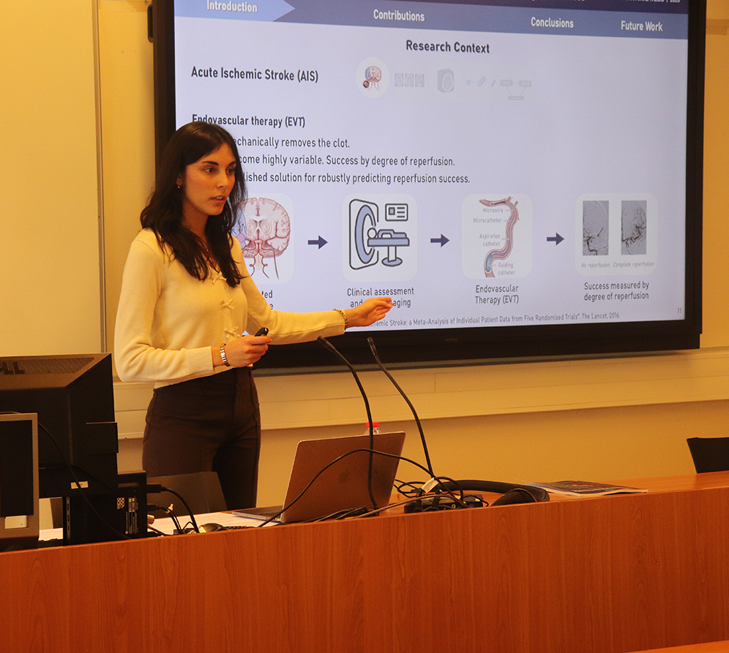
This PhD thesis focuses on the development of deep learning methods to enhance medical image analysis, with a particular emphasis on improving focus, efficiency and fairness in acute ischemic stroke (AIS). AIS is a cerebrovascular disease that occurs when a cerebral artery becomes occluded, interrupting blood flow to part of the brain. Rapid diagnosis and treatment of AIS are essential to preserving salvageable tissue and minimizing long-term disability. However, not all patients benefit equally from current therapies despite similar clinical and procedural characteristics. Accurately identifying patients likely to benefit remains a major clinical challenge. In this context, neuroimaging, particularly brain Computed Tomography Angiography (CTA), plays a central role in patient triage during the acute stroke phase. The rich anatomical and vascular information present in CTA offers a valuable but complex input for automated image analysis, motivating the use of deep learning to support clinical decision-making.
This thesis explores strategies to fully utilize CTA data using deep learning methods tailored for AIS. The first contribution proposes strategies to guide neural networks toward vascular structures extracted from CTA while retaining contextual parenchymal information. Several methods to combine vascular segmentations and CTA data are evaluated, including an attention-inspired mechanism designed to enhance the model’s focus on clinically relevant features for AIS due to large vessel occlusion (LVO) detection. The second contribution introduces Learnable 3D Pooling (L3P), a novel convolutional neural network-based module that compresses
3D medical images into 2D feature maps, enabling efficient, lightweight, and interpretable models. L3P-based architectures are validated across multiple tasks, including LVO detection in CTA, as well as brain age prediction from 3D T1-weighted MRI. In all cases, L3P maintains competitive performance compared to fully 3D networks, while significantly reducing computational demands and enhancing feature interpretability. The third contribution addresses the issue of hidden confounders in medical imaging pipelines. A controlled experimental framework is proposed to simulate and analyze the effects of confounding variables in clinically relevant classification tasks. Using large ensembles of bootstrapped models, measurable distributional patterns, such as inflated model performance, reduced performance variability and convergence of training and validation metrics, are identified as indicators of confounder-driven learning. This enables the development of a practical, unsupervised method to flag potential hidden biases, even when the nature of the confounder is unknown.
The proposed approaches are broadly applicable across medical imaging modalities and disease domains, with particular relevance to neuroimaging. Designed with generalizability in mind, these methods have demonstrated utility beyond stroke, highlighting their potential for diverse clinical contexts. Collectively, the contributions of this thesis advance the development of deep learning tools with efficiency, focus, and fairness.
The complete doctoral thesis can be consulted in the official PhD repository of the University of Girona (DUGi-doc): https://dugi-doc.udg.edu


